Cochlear implants are suitable for patients with a severe to profound sensorineural hearing loss, but what does that mean? To understand this we first need to describe the anatomy of the ear. The ear has three main parts: the outer ear, the middle ear and the inner ear. Click on the sections below to find out more about each part;
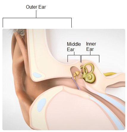
The Outer Ear (Pinna and Ear Canal)
The bit of our ears that we can see on the outside of our head, is called the pinna or auricle. It is there not just to hold our glasses on or add earrings to, it collects and channels sound into the ear canal. The hole that we can see in our ears is called the ear canal (also known as the external auditory meatus). This directs the sound into the ear and has glands that produce wax. Although sometimes a nuisance, wax does a brilliant job at keeping our ears clean and free from debris.
The Middle Ear (Tympanic Membrane and Ossicular Chain)
The eardrum is also known as the tympanic membrane. When sound travels down the ear canal it reaches the ear drum and changes the sound into vibrations. The ossicular chain is made up of the hammer, anvil and stirrup (also known as the malleus, incus and stapes). This chain of three small bones (the smallest in your body), transfers and amplifies the vibrations from the ear drum into the inner ear. The stapes footplate hits onto the membrane of the round window in the cochlear. There is also a muscle attached to the ossicular chain that can reduce movement when there is a loud noise, in order to help prevent damage to the inner ear.
The Inner Ear
The inner ear (cochlea) contains the basilar membrane surrounded by fluid. Along the membrane are highly sensitive hair cells. When the basilar membrane is moved by the surrounding fluid, these tiny hair-like structures move, a neurotransmitter is released and the auditory nerve is then stimulated. See below for a video clip of a hair cell responding to music making the basilar membrane move. The hair cells are responsible for converting the vibration into an electrical signal that can be passed onto the auditory nerve and taken up to the brain. The vestibular system is also found in the inner ear. The semicircular canals of the inner ear are responsible for giving information to help us balance.
Inner hair cells in the cochlea
As mentioned above, hair cells change sound vibrations into electrical signals that are sent to the brain via the hearing nerve. The link to the video below is a demonstration of a hair cell being stimulated by music. Click on the link to watch the video.
https://www.youtube.com/watch?v=wKEhjpqpzCE
Key Concepts